Discussion
Loading...

Post

  • About
  • Code of conduct
  • Privacy
  • Users
  • Instances
  • About Bonfire
RoedigerRG
@RoedigerRG@social.anoxinon.de  ·  activity timestamp 4 days ago

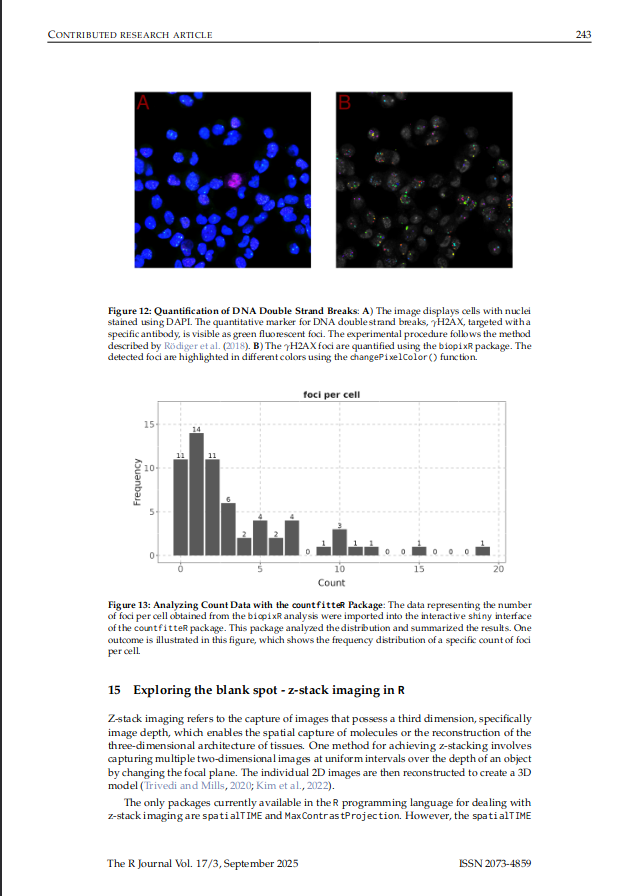
📢 @devTLB and I’ve just published a review article titled **“Exploring Image Analysis in R: Applications and Advancements”** in *The R Journal* (Vol. 17, Issue 3).

🔍 The paper surveys #rstats image‑analysis packages, from the early work of *EBImage* to the newer packages like *biopixR*. The majority of these packages are still active, with two‑thirds being updated. A robust and vibrant ecosystem for bioimage informatics.

🔗 You can read the full article here: doi.org/10.32614/RJ-2025-030

 Image from 'Exploring Image Analysis in R' by Brauckhoff et al. The image contains two parts: Figure 12 and Figure 13. Figure 12 displays two fluorescence microscopy images of cells with stained nuclei. In image A, the nuclei are blue, and DNA double-strand breaks (H2AX foci) are highlighted in red. Image B shows a similar view but with different color highlighting. Figure 13 is a bar graph showing the frequency distribution of gamma H2AX foci counts per cell. The x-axis represents foci count (0 to 20), and the y-axis represents frequency.
Image from 'Exploring Image Analysis in R' by Brauckhoff et al. The image contains two parts: Figure 12 and Figure 13. Figure 12 displays two fluorescence microscopy images of cells with stained nuclei. In image A, the nuclei are blue, and DNA double-strand breaks (H2AX foci) are highlighted in red. Image B shows a similar view but with different color highlighting. Figure 13 is a bar graph showing the frequency distribution of gamma H2AX foci counts per cell. The x-axis represents foci count (0 to 20), and the y-axis represents frequency.
  • Copy link
  • Flag this post
  • Block
Log in

Open Science

We are a network of scientists, developers and organizations building the next generation of digital spaces for open science.

Open Science: About · Code of conduct · Privacy · Users · Instances
Bonfire open science · 1.0.0-rc.4.1 no JS en
Manual federation enabled
  • Explore
  • About
  • Members
  • Code of Conduct
Home
Login