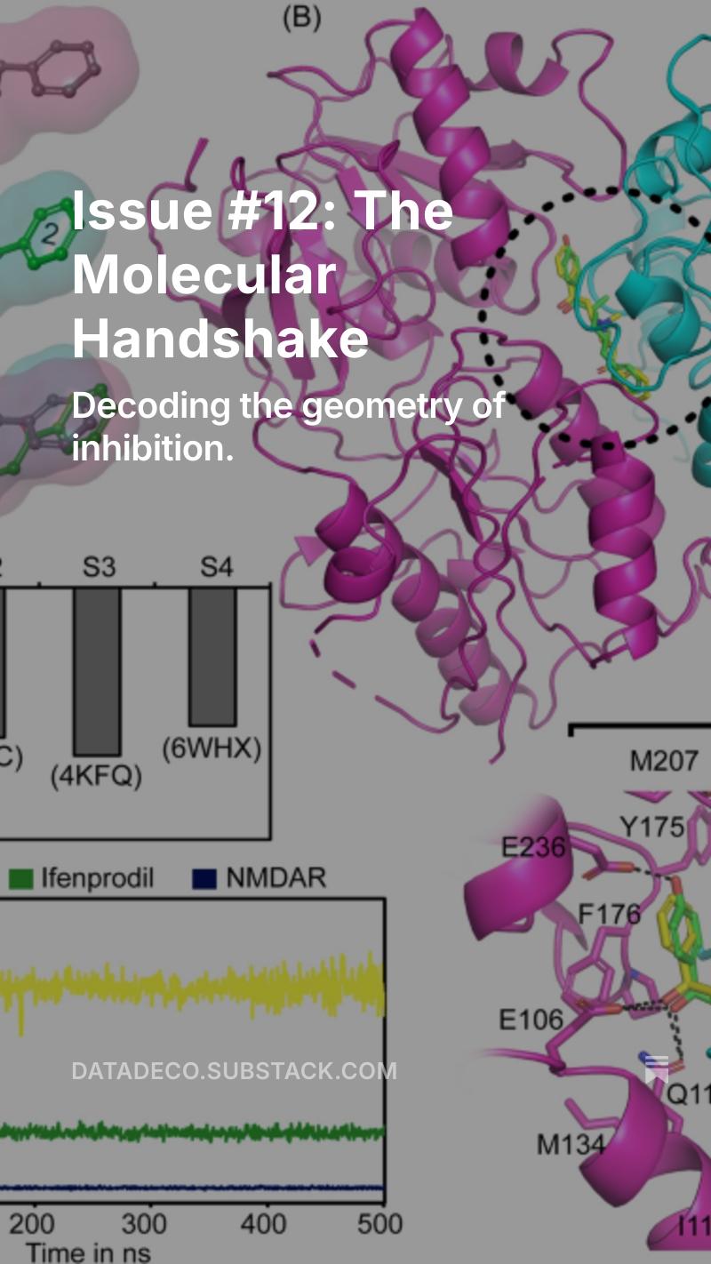
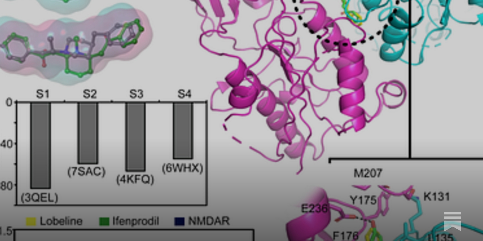
A scientific figure showing molecular binding. Panel A displays the chemical structures of ifenprodil and lobeline in pink and green translucent bubbles. Panel B features a large, colorful 3D model of the NMDAR receptor made of magenta and cyan ribbon-like spirals, with small yellow and green molecules bound at their interface. Panels C and D show grey bar graphs and a yellow-and-green line graph representing binding energy and molecular stability over time.