Discussion
Loading...

Post

Log in
  • About
  • Code of conduct
  • Privacy
  • Users
  • Instances
  • About Bonfire
Stefan Rödiger
Stefan Rödiger
@pangolin  ·  activity timestamp 4 months ago

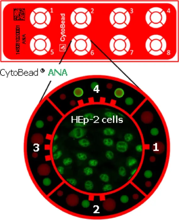
Our new study evaluates the performance of the CytoBead ANA 2 assay, a novel one-step method for detecting antinuclear antibodies (ANA) and specific autoantibodies in systemic autoimmune rheumatic diseases (SARDs).


Roei, T., Boris, G., Yehuda, Shovman, Milena, T., Eleanor, Z., Ariel, B., Tima, D., Büttner, T., Nasser, A., Michel, J., Rödiger, S., Schierack, P., Howard, A., Roggenbuck, D., Yehuda, Shoenfeld, Ora, S., 2025. CytoBead ANA 2 assay - a novel method for the detection of antinuclear antibodies. Sci Rep 15, 22131. https://doi.org/10.1038/s41598-025-04583-3


We compared CytoBead ANA 2 to traditional ANA indirect immunofluorescence (IIF) and the BioPlex™ 2200 multiplexed assay, finding substantial agreement (κ=0.74) and comparable diagnostic accuracy. The assay also effectively identified the dense-fine speckled (DFS) pattern associated with anti-DFS70 antibodies in non-SARDs patients.

This approach streamlines ANA testing by combining screening and confirmation in a single step, offering a promising alternative to the current two-tier process.

Read the full paper: Nature Scientific Reports

#Autoimmunity #Rheumatology #Diagnostics #ANA #Research

CytoBead ANA 2 Slide Format

This image illustrates the CytoBead ANA 2 slide format, designed for the simultaneous screening and confirmation of antinuclear antibodies (ANA) and ANA-specific autoantibodies.

- Central Compartment: Contains HEp-2 cells, which are used for the initial screening of ANA via indirect immunofluorescence. The presence of autoantibodies is indicated by fluorescence patterns on these cells.
- Peripheral Compartments: Surrounding the central HEp-2 cell compartment are four smaller compartments, each containing antigen-coated fluorescent microbeads. These compartments are used for the confirmatory detection of specific autoantibodies:

- Compartment 1: Contains SS-B/La-coated beads (9 µm) and Jo-1-coated beads (15 µm).
- Compartment 2: Contains Sm-coated beads (9 µm) and Sm/RNP-coated beads (15 µm).
- Compartment 3: Contains dsDNA-coated beads (9 µm) and Scl-70-coated beads (15 µm).
- Compartment 4: Contains SS-A/Ro60-coated beads (9 µm) and SS-A/Ro52-coated beads (15 µm).

Reference Beads: Size and location reference (SLR) beads are present in all peripheral compartments to assist in the identification and localization of the antigen-coated microbeads.

This layout allows for both the screening of ANA on HEp-2 cells and the simultaneous confirmation of specific autoantibodies using antigen-coated microbeads, streamlining the diagnostic process.
CytoBead ANA 2 Slide Format This image illustrates the CytoBead ANA 2 slide format, designed for the simultaneous screening and confirmation of antinuclear antibodies (ANA) and ANA-specific autoantibodies. - Central Compartment: Contains HEp-2 cells, which are used for the initial screening of ANA via indirect immunofluorescence. The presence of autoantibodies is indicated by fluorescence patterns on these cells. - Peripheral Compartments: Surrounding the central HEp-2 cell compartment are four smaller compartments, each containing antigen-coated fluorescent microbeads. These compartments are used for the confirmatory detection of specific autoantibodies: - Compartment 1: Contains SS-B/La-coated beads (9 µm) and Jo-1-coated beads (15 µm). - Compartment 2: Contains Sm-coated beads (9 µm) and Sm/RNP-coated beads (15 µm). - Compartment 3: Contains dsDNA-coated beads (9 µm) and Scl-70-coated beads (15 µm). - Compartment 4: Contains SS-A/Ro60-coated beads (9 µm) and SS-A/Ro52-coated beads (15 µm). Reference Beads: Size and location reference (SLR) beads are present in all peripheral compartments to assist in the identification and localization of the antigen-coated microbeads. This layout allows for both the screening of ANA on HEp-2 cells and the simultaneous confirmation of specific autoantibodies using antigen-coated microbeads, streamlining the diagnostic process.
  • Copy link
  • Flag this post
  • Block

Open Science

We are a network of scientists, developers and organizations building the next generation of digital spaces for open science.

Open Science: About · Code of conduct · Privacy · Users · Instances
Bonfire open science · 1.0.1 no JS en
Manual federation enabled
Log in
  • Explore
  • About
  • Members
  • Code of Conduct